
This video opens up the inside of cells, in a way never seen before.
The research published in Cell uses new microscopy techniques to explore, in 3D, tiny structures inside cells such as mitochondria, the "fuel pack" of a cell.
The researchers argue that their approach “permits ultra-high resolution 3D imaging of virtually any subcellular structure”. They hope this will allow previously unanswerable questions about cell biology to be tackled, potentially opening up new avenues in curing disease.
The images of a mouse cell are made of millions of molecules, which appear as bright white dots, before turning into coloured dots. They represent structures that pair paternal and maternal chromosomes during reproduction.
As the commentary highlights, the video allows an extraordinary new level of detail. The researchers have also mapped a number of other structures, as shown in this graphic.
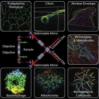
The researchers say that imaging a range of organelles (tiny structures within cells that perform specific functions) is a powerful tool. It “promises a new perspective on how proteins distribute across entire organelles throughout whole cells, a key unmet challenge in cell biology”.
Have you read?